

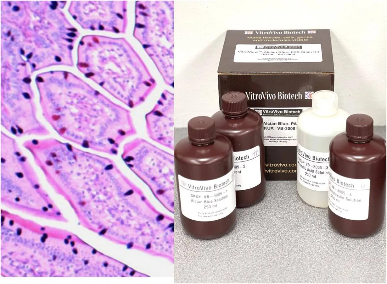
Mucosal tissues are dynamic structures where secreted material, epithelial cells, glands, vessels, and supporting connective tissue interact closely. An alcian blue pas stain can help researchers distinguish acidic and neutral mucins, providing a clearer view of how secretory composition varies across a tissue section.
That chemical information becomes more useful when researchers also examine the framework supporting those cells. Looking at mucins and stromal architecture as separate but related features can reveal changes that routine morphology alone may not show clearly.
Begin With the Secretory Compartment
Mucins contribute to protective and lubricating surfaces in organs such as the respiratory and gastrointestinal tracts. Changes in their amount or composition may accompany experimental injury, inflammation, differentiation, or altered glandular activity, making selective histochemistry useful in tissue-based research.
A Reticulin stain kit addresses a different part of the same tissue environment by highlighting reticular fibers. These delicate fibers form supportive networks around cells and tissue compartments, allowing investigators to examine stromal organization alongside epithelial or glandular changes.
Understand the Alcian Blue–PAS Pattern
The combined method uses Alcian Blue to demonstrate acidic mucins and the PAS reaction to show neutral mucins in a contrasting color. Acidic mucins appear blue, neutral mucins appear magenta, and mixtures can produce blue-purple regions.
This color separation can help researchers map secretory differences across glands, epithelial surfaces, or defined regions of interest. The result should still be interpreted with morphology, because staining intensity alone does not explain why mucin composition has changed.
Read the Supporting Fiber Network
Reticular fibers are fine components of connective tissue that help organize cellular and stromal compartments. Silver-based reticulin staining makes these fibers dark and easier to follow against a contrasting background, supporting structural assessment across selected tissue regions.
Changes in fiber continuity, density, or arrangement may provide context for epithelial remodeling or altered tissue organization. Researchers should avoid treating reticulin changes as a direct measure of mucin biology; the two stains answer different questions and should be interpreted separately first.
Use Adjacent Sections Strategically
Adjacent sections can make it easier to compare secretion patterns with supporting architecture. One slide may be stained for acidic and neutral mucins, while a nearby section is reserved for reticular fibers under conditions optimized for the silver-based method.
Because consecutive sections are not identical, researchers should record cutting level, orientation, and anatomical landmarks. This is especially important in small glands or focal lesions where a structure may shift noticeably between neighboring sections.
Keep Processing Conditions Consistent
Fixation, section thickness, deparaffinization, hydration, reagent timing, differentiation, and mounting can all influence final staining quality. Standardizing these variables reduces the chance that technical differences will be mistaken for biological variation between experimental groups.
The Alcian Blue–PAS workflow also requires careful sequencing so acidic and neutral mucins remain distinguishable. Reticulin staining has its own oxidation, silver impregnation, reduction, and counterstaining requirements, so each protocol should be optimized independently.
Build Controls Into the Study
Positive control tissue can confirm that the expected color or fiber reaction is developing correctly. Controls are particularly helpful when new tissue types, multiple batches, or archived samples are involved, because they provide a stable reference for staining performance.
Researchers should process controls alongside experimental slides whenever practical. Recording reagent lots, incubation times, section thickness, and deviations from standard procedures can also make troubleshooting faster when an unexpected staining pattern appears.
Standardize Imaging and Scoring
Microscope settings should remain consistent when images will be compared across samples. Magnification, illumination, exposure, white balance, and digital processing can alter the apparent intensity of blue, magenta, or dark fiber staining.
Scoring criteria should also be established in advance. Researchers may assess mucin distribution, positive area, gland-specific patterns, fiber density, or structural continuity, but the same region-selection and measurement rules should be applied throughout the study.
Interpret Structure and Secretion Together
Mucin composition and reticular architecture may change in the same specimen without changing in parallel. A tissue can show altered secretion while retaining its supporting framework, or structural remodeling can occur without a major shift in mucin pattern.
Treating each endpoint independently first prevents overinterpretation. Researchers can then compare the results with H&E morphology, experimental treatment, time point, or molecular data to understand how secretory and stromal changes relate within the broader tissue response.
Conclusion
Mucosal tissue research benefits from methods that examine both what cells produce and how the surrounding tissue is organized. Mucin-focused histochemistry and reticular-fiber staining provide complementary information, but each requires its own preparation, controls, and interpretation strategy.
By planning adjacent sections, standardizing processing, defining scoring rules, and maintaining consistent imaging, researchers can extract more information from limited tissue. This coordinated approach creates a clearer picture of how secretory composition and stromal architecture change across experimental conditions.
